

Truly painless cancer treatment, realized through medical image analysis.
4D-CT generation brings respiratory tumor motion into the treatment plan, supporting precise delivery to moving tumors.
Generating 4DCT images from 3DCT images
Artificial Intelligence
Radiation therapy and particle therapy have the potential to benefit from the use of 4D CT imaging. However, there are several challenges that need to be addressed, such as artifacts caused by irregular breathing patterns and high radiation exposure associated with 4D CT scans. That is why we have developed a product that uses our Artificial Intelligence technology to generate high-precision 4D CT images from 3D CT images. This enables more accurate and safer treatment, providing peace of mind for medical staff.

Higher accuracy of treatment planning
The thoracoabdominal region is highly affected by respiratory motion, and in particle therapy, density changes can have a significant impact on dose distribution. To address these issues, various techniques such as 4D robust optimization and range-adaptive ITV have been developed, but these require 4D CT images. However, even with only 3D CT images, our product can generate 4D CT images, enabling more accurate treatment planning and potentially improving treatment outcomes.

Application expansion of machine learning + IGRT
In the medical field, precise treatment is essential. To achieve this, image-guided radiation therapy (IGRT), which accurately identifies the position of a tumor that moves with respiration and irradiates it with a treatment beam, has become the mainstream. However, the structures within the body are complex, and simple image processing techniques often fail to achieve sufficient accuracy. Therefore, IGRT using machine learning has been gaining attention recently. With our product, even with 3D CT images, high-precision machine learning IGRT can be achieved by using them as training data. This enables medical staff to provide more accurate treatments that meet their needs.
How 4DCT image generation works
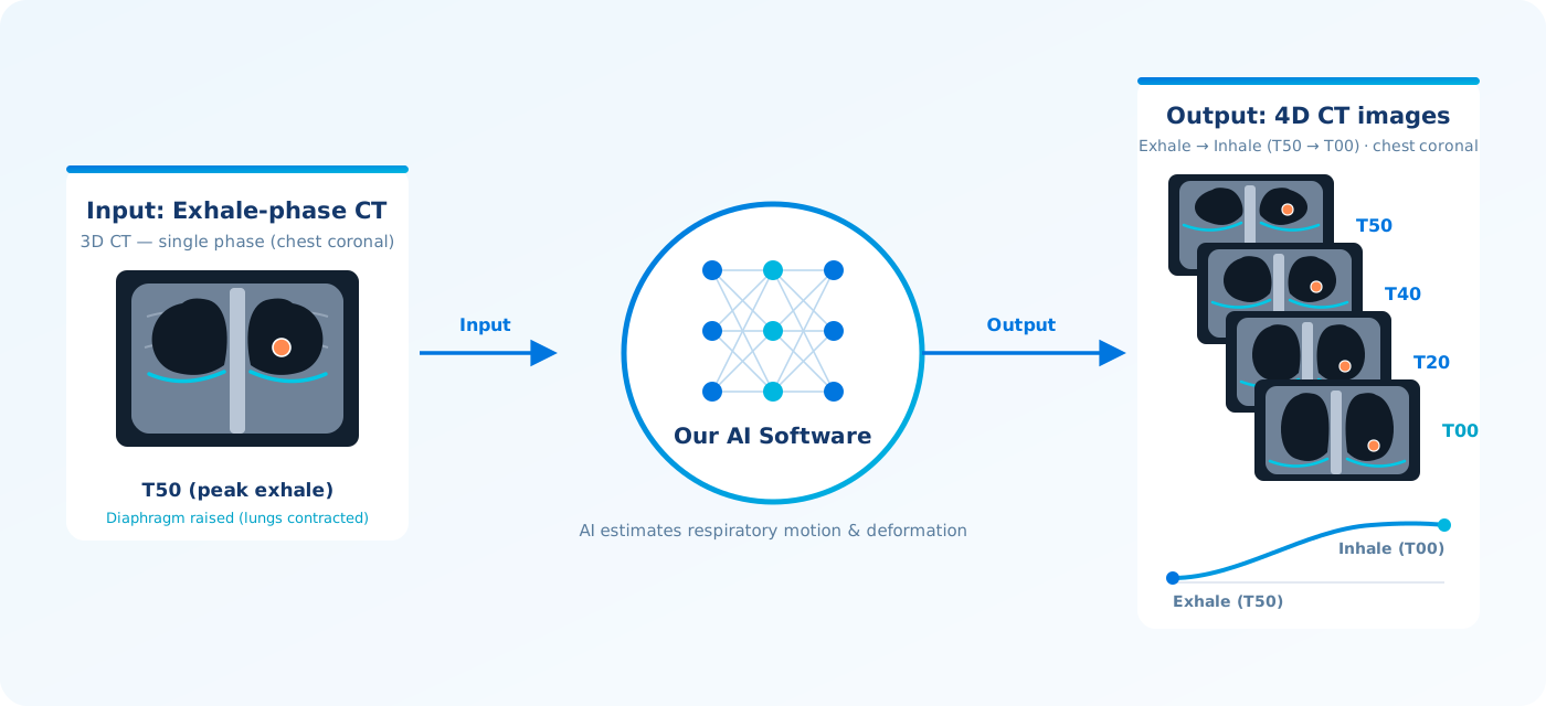
Simply input a 3D CT image acquired at the exhale phase into our software, and the AI estimates organ motion and deformation caused by respiration, automatically generating and outputting CT image data for every respiratory phase from exhale to inhale. Since no actual 4D CT scan is required, radiation dose can be reduced and artifacts caused by irregular breathing patterns can be avoided. The generated 4D CT image data can be used directly for treatment planning and as training data for image-guided radiation therapy (IGRT).
Clinical example: generated 4D-CT images
Data source / citation: Hugo, G. D., Weiss, E., Sleeman, W. C., Balik, S., Keall, P. J., Lu, J., & Williamson, J. F. (2016). Data from 4D Lung Imaging of NSCLC Patients (Version 2) [Data set]. The Cancer Imaging Archive. https://doi.org/10.7937/K9/TCIA.2016.ELN8YGLE
License: used under CC BY 3.0.
Modification notice: 4D-CT images were generated by our software from the exhale-phase CT image in the TCIA data set; cross-sections were extracted, windowed and rendered as a video for display.
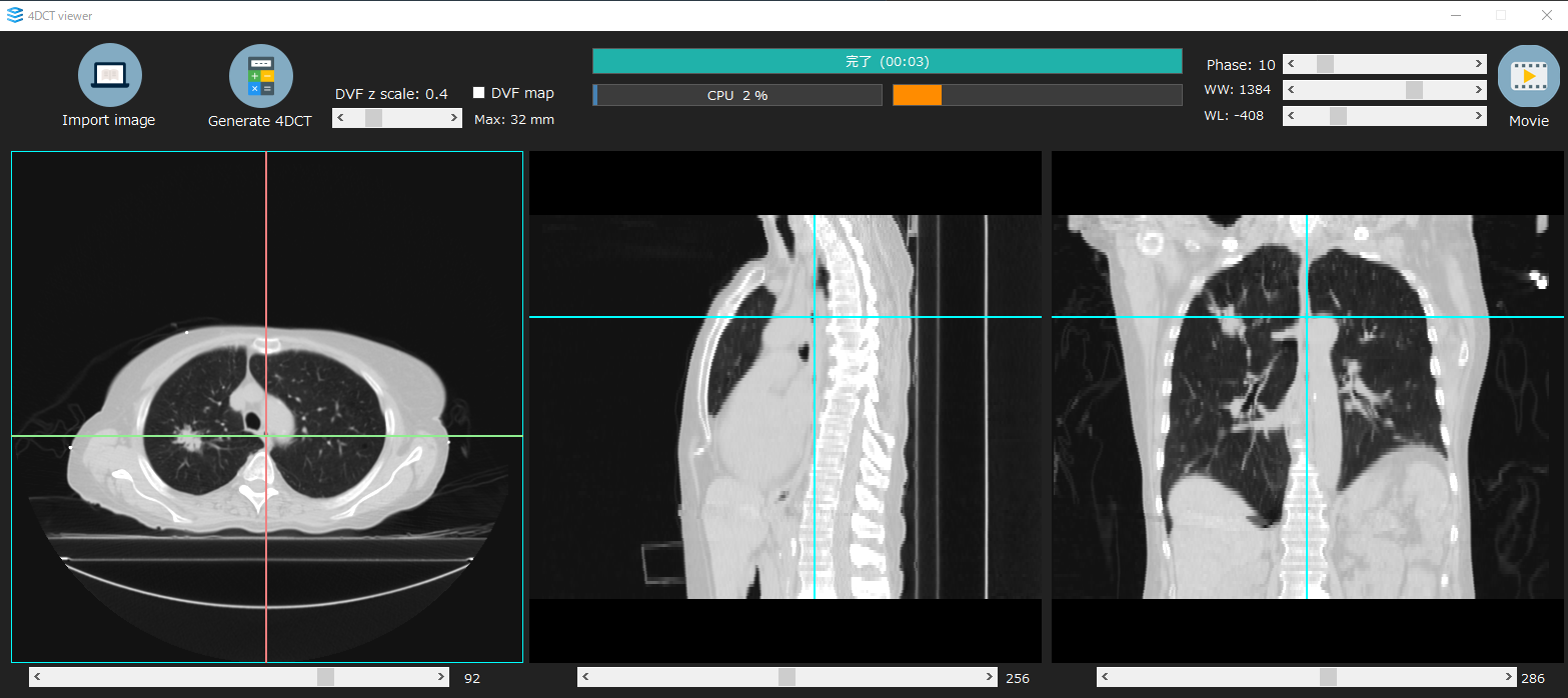
A simple viewer is included
- Everything from image import to 4D-CT generation, phase display and movie export runs from batch files; no dedicated installation is required.
- Axial, sagittal and coronal views are displayed in sync, with sliders for respiratory phase and window width / level.
- The respiratory motion amplitude (DVF scale) can be adjusted manually, so the 4D-CT can be matched to each patient's breathing.
Contact
Let's solve medical imaging,
together.
Joint research, product development support, careers — feel free to get in touch.